Monday, 9 March 2020

DVT / PE

What symptoms would a patient complain of if they had a DVT? 

Pain (usually unilateral calf) SIN = severe!
*When you see a lot of orthopaedic surgery like TKR, THR you get a good feel for what is the typical amount of pain. In my experience people with DVT have a profound level of pain.

S = Severe
I = High
N = Inflammatory (cardinal signs dolor, tumor, rubor,
Quality = Heavy ache

Clinical point: 
Always ask questions and examine a suspected DVT with suspicion of PE
e.g. Any SOB, chest pains, low SATS?

OBS: Dilated superficial veins, calf redness, discolouration, lower limb swelling
PALPATION: Pitting oedema, tenderness and firmness on palpation of the calf
SPECIAL TESTS: +ve Homan’s Sign

What would you do if a patient you were seeing presented with these symptoms?

Inpatient on the ward:
Escalate to nurse / RMO / consultant

Outpatient: 
Urgent GP referral for further Ix
A+E if suspected PE

Patients can be divided into “DVT unlikely” and “DVT likely” groups based on Wells score. An additional moderate risk group can be added based on the sensitivity of the d-dimer being used.
  • A score of 0 or lower is associated with DVT unlikely with a prevalence of DVT of 5%.
    • These patients should proceed to d-dimer testing:
      • A negative high or moderate sensitivity d-dimer results in a probability <1 and="" further="" imaging="" is="" li="" no="" required.="">
      • A positive d-dimer should proceed to US testing.
        • A negative US is sufficient for DVT rule out.
        • A positive US is concerning for DVT; strongly consider treatment with anticoagulation.
  • A score of 1-2 is considered moderate risk with a pretest probability of 17%.*
    • These patients should proceed to high-sensitivity d-dimer testing (moderate sensitivity d-dimer is not sufficient).
      • A negative high-sensitivity d-dimer is sufficient for rule out of DVT in a moderate risk patient with a probability of <1 li="">
      • A positive high sensitivity d-dimer should proceed to US testing.
        • A negative US is sufficient for ruling out DVT.
        • A positive US is concerning for DVT, strongly consider treatment with anticoagulation.
  • A score of 3 or higher suggests DVT is likely. Pretest probability 17-53%.
    • All DVT likely patients should receive a diagnostic US.
    • D-dimer testing should be utilized to help risk-stratify these DVT-likely patients.
      • In DVT likely patients with negative d-dimer:
        • A negative US is sufficient for ruling out DVT, consider discharge.
        • A positive US should be concerning for DVT, strongly consider treatment with anticoagulation.
      • In DVT likely patients with a positive d-dimer:
        • A positive US should be concerning for DVT, strongly consider treatment with anticoagulation.
        • A negative US is still concerning for DVT. A repeat US should be performed within 1 week for re-evaluation.


Tuesday, 17 January 2017

Red Flags / Yellow Flags

Q. What are they? 

A. Signs and symptoms that indicate serious spinal pathology 


  • Age > 55 (above this age, particularly above 65) the chances of being diagnosed with serious pathologies such as cancer increases

  • PMHx or FHx of Ca  

  • Unexplained weight loss (5% within a 4 week period not related to change in diet or exercise) 

  • Constant / unremitting / progressive / non-mechanical pain (does not vary in a 24 hr period) 

  • Night pain (due to increase bony lysis at night) 

  • Generally unwell (fever / night sweats / signs of systemic illness) 

  • Bilateral neurological signs 

  • Ataxic gait (lack of muscle coordination / uncoordinated / jerky movements / poor balance) could indicate MS, stroke or TIA, head trauma, damage to cerebellum, spinal cord damage, brain tumour)

  • Major trauma (fall from a height / RTA) 

  • Minor trauma (in osteoporotic patients) 

  • Thoracic pain (most common region for metastases) 

  • Long term use of steroids 

Cauda Equina (require immediate referral to hospital for neurosurgical) 


  • Saddle anaesthesia (perianal sensory loss) 

  • Bladder or bowel dysfunction (urinary retention / incontinence or faecal incontinence) 

  • Bilateral leg pain / sciatica / neurological signs 

Yellow Flags 


Q. What are they? 

A. Bio-psychosocial factors that may risk increase likelihood of chronicity 

  • Pending compensation claim 
  • Off word 
  • Low work status 
  • Anxiety 
  • Depression 
  • Inappropriate coping strategies 
  • Passive dependent locus of control 

A - Attitudes 

Towards the current problem. Does the patient feel that with appropriate help and self management they will return to normal activities?

B - Beliefs 

The most common misguided belief is that the patient feels they have something serious causing their problem-usually cancer. 'Faulty' beliefs can lead to catastrophisation.

C - Compensation 

Is the patient awaiting payment for an accident/ injury at work/ RTA?

D - Diagnosis 

or more importantly Iatrogenesis. Inappropriate communication can lead to patients misunderstanding what is meant, the most common examples being 'your disc has popped out' or 'your spine is crumbling'.

E - Emotions

Patients with other emotional difficulties such as ongoing depression and/or anxietous states are at a high risk of developing chronic pain.

F - Family 

There tends to be two problems with families, either over bearing or under supportive.

W - Work

The worse the relationship, the more likely they are to develop chronic LBP.

Common Anterior Hip Complaints DDx / Assessment / Treatment

Credit to most of this goes to Nichole Hamilton with some additional thoughts from myself

"Carly Says Jacket Off"


Cartilage: Labral tear / articular cartilage wear

Soft tissue: Anterior capsule sprain, Iliopsoas tendinopathy, Iliopsoas bursa

Joint: OA / Synovitis

Other Dx: Stress # (NOF, Pubic Ramus), Osteitis Pubis, Hernia, Abdominal Pathology

"WHY is the anterior hip painful?"

Excessive load on the anterior hip can occur due to: 

1) Structural (bony) 


FAI:  CAM, PINCER or combined 

2) Biomechanical dysfunction 

Increased biomechanical load on the anterior hip with excessive anterior translation of femoral head (Sahrmann, 2003) 

1) Postural Alignment 

- Standing with hips in extension (watch out for sway back postures) 
- Genu recurvatum (knee hyperextension) 

"Want all the floors stacked on top of each other" 

Plumb line to align the femoral head in the roof of the acetabulum... 

Greater trochanter in line with...

Lateral femoral condyle and... 

Just anterior to lateral malleolus 

ASIS like car headlights facing dead ahead 

Manu-sternal junction over pubic symph 

2) Hypertonicity in deep posterior hip muscles / Tight Posterior Capsule 

Much like the shoulder if the posterior cuff is tight gets tight, develops trigger points, these muscles blend with the posterior capsule and prevent posterior glide (squeezes it anteriorly) 

Femoral head will follow the path of least resistance and if tight posteriorly will glide anteriorly and cause impingement 

Why do these posterior hip muscles get tight? Potentially more motor control strategies in LPHC, tightness compensating for poor stability somewhere else, poor lumbar-pelvic stability. 

3) Long / inhibited psoas 

A dynamic restraint that crosses the anterior aspect of the hip joint before it inserts into the lesser trochanter. Can protectively tighten to prevent / protect against shear and subluxation. 

Stretching in a hyperextended position with an anterior pelvic tilt can loosen the anterior capsule / iliofemoral ligament and cause this protective tightnening and can lead to labral stress. 

2 Pathways to Anterior Hip Impingement / Labral Tear 

Long inhibited psoas leads to... 
Reduced hip stability and motor control... 
Increased anterior translation of femoral head in extension (standing, running, wearing high heels)...
Increased load on anterior hip structures

OR 

Articular SIJ or lumbar dysfunction leads to...
Poor motor control strategies... 
Hypertonic posterior hip...
Restricted posterior glide during flexion (squatting for example)...
Anterior hip impingement with flexion

BOTH MAY RESULT IN... 

Anterior impingement / labral tear 

4) Hamstring Dominant Hip Extension 

Hamstrings being used as primary hip extensions mean that the axis of movement occurs at the origin (ischial tube) below acetabulum. The distal part of femur heads into extension but the femoral head anteriorly. Glutes cross the hip joint and don't produce that anterior translation. 

Watch out of hamstring synergistic dominance (performing movements in an anterior pelvic tilt e.g. squat or deadlift) alters length-tension relationship of glutes and puts them at a mechanical disadvantage, more likely for hammys to become primary hip extensor. 

5) Low Back 

Patients with CLBP...

Increased tonic activation of glutes in patients during low load tasks e.g. standing (glutes should be firing phasically) 

Also showed to have poor phasic strength, delayed firing (e.g. during an activity like climbing the stairs) 

Reduced cross-sectional area of psoas in patients with unilateral back pain 

During ASLR reduced activity of psoas results in increased anterior hip joint force (Lewis & Sahrmann, 2009) 

Also check ipsilateral SGIS 

6) Foot / ankle 

Inability to pronate (shock absorb) and supinate 

Excessive rigidity in forefoot, mid-foot, rear-foot can contribute to anterior hip load. 

Reduced ability to shock absorb. 

More likely to have hip in externally rotated position if stuck in supination 

Watch and listen to someone doing single leg hops (from one foot to the other) does it sound louder one side vs the other? i.e. poor pronation / shock absorption

7) Thorax  

One of the rotational centres (along with the hip) 

Asymmetry in thoracic rotation 

Should be equal range L vs R 

If limited L for example thorax will tend to sit in mid range (pointing slightly right) 

Hips with rotate left to have ASIS pointing forward and position right head of femur anteriorly in the acetabulum 

8) Anterior core control 

Able to prevent anterior tilt during hip flexion based activity e.g. squatting 

Assessment 

Subjective: When does the patient feel the symptoms? Flexion or extension? 

Flexion intolerant? 

Extension intolerant?

Labral tear: Insidious onset (rarely traumatic) groin pain related to hip movement usually reproduced with rotational movement e.g. pivoting / twisting, clicking/catching sensation often reported. 

Test: Does femoral head remained centered during the painful activity?

Special tests: Quadrant test / FADIR looking for reproduced of symptoms 

Observe: Are there any other areas in the chain that might be adding to the load on the anterior hip? 

Foot, thorax, SIJ? 

Palpate: Any local areas of the hip that are contributing to poor hip centering? 

Treatment 


Educate: Posture, daily habits, hip mechanics, pain education 

Manual therapy: Areas of hypertonicity that might be contributing to non-centering of femoral head (TFL, rec fem, piriformis, OI, posterior pelvic floor which has links to obturator internus). Foot, thorax, SIJ, lumbar. 

Exercises: Self release, correct core activation, functional hip centering, postural alignment, strengthening (lower fibres glute max, GMed, quadratus femoris, core stability, closed chain exercises e.g. bridge vs open chain hip extension. 

Avoid hip stretches (psoas, piriformis) can cause more harm than good 

Imagining: MRI or MR Arthrogram 

Surgery: Traumatic injury (20-30%) tend to be people who benefit best from surgery (not so much a biomechanical dysfunction causing labral tear), physio mx unsuccessful (3months no improvement), surgery often not essential for every patient.

Patients with OA signs in hip don't tend to do as well with hip arthroscopy


Thursday, 31 October 2013

Predisposing factors to cervical instability

Factors that can predispose to cervical instability: 

Lax posterior transverse ligament (joining the dens to C1) thought to be a risk factor for atlantoaxial instability (Merrick et al, 2000) 


  • Main movement occurring at this level (atlantoaxial jt) is rotation of 35 degrees in each direction
  • Rotation is limited by the alar ligaments 


Pathologies 

Systemic inflammatory diseases (because the inflammation damages connective tissue, ligaments, joints etc) 


  • Lupus
  • RA (most common in cervical spine and MCP joints in the hands)
  • Ankylosing spondylitis (primarily affects c-spine)
  • Down Syndrome (laxity or congenital absence of transverse atlas ligament which can make minor traumas to the neck able to cause subluxation)


  • Degenerative changes of cervical spine (Andrews, 1981) 
  • Hypermobility 
  • Connective tissue disorders (Ehler's-Danlos syndrome, Marfan syndrome) 
  • Congenital bony abnormalities e.g. short dens, odontoid hypoplasia (basically geeky talk for the same thing!) 


PMH

Previous or recent RTAs (recurrent whiplash) 
OA 
General wear and tear 

DH (medications) 

Long term use of steroids 

Metastatic Disease 

Most prevalent in thoracic spine (1st) lung and breast primary tumors 

Lumbar (2nd) Prostate, colorectal, ovarian cancers 

Cervical (3rd) 


Red Flags of metastatic disease 

Non-mechanical picture (pain not affected by positional changes) 

Unremitting pain (no diurnal or nocturnal variation) 

Night pain and pain worse at night (this is due to more bony lysis occuring aka breaking down or erosion of the bone) 

References 

Merrick J, Ezra E, Josef B, Hendel D, Steinberg DM, Wientroub S. Musculoskeletal problems in Down Syndrome European Paediatric Orthopaedic Society Survey: the Israeli sample. J Pediatr Orthop B. Jun 2000;9(3):185-92.

Andrews LG. Myelopathy due to atlanto-axial dislocation in a patient with Down's syndrome and rheumatoid arthritis. Dev Med Child Neurol. Jun 1981;23(3):356-60.

Friday, 11 October 2013

Morphine OD

I was watching the film "Prisoners" where at the end a little girl is given a lethal dosage of morphine. My flat mate Ed (a med student) told me that the first thing that would kill you from a morphine OD is respiratory failure. As your rate and depth of breathing reduces eventually it's not enough to keep you alive.

The medical fix is naloxone, a drug used to counter the effects of an opiate overdose. BUT the half life of naloxone is much shorter than morphine, it's broken down quicker so patients should be kept in for at least 24 hrs for observation.

Sunday, 15 July 2012

Ehlers–Danlos syndrome

A group of inherited connective tissue disorders caused by a defect in the synthesis of type I or type III collagen.

S&S 

MSK


  • Hypermobile joints 
  • Hx of sprains, subluxation, dislocations 
  • Swan neck deformity 
  • Weak muscle tone 
  • Osteopenia

Wednesday, 16 May 2012

Knee extension test in sitting

Testing if a patient can actively extend both knees and achieve at least a 3/5 grade knee extensor strength on Oxford scale is a basic way of assessing their capacity to attempt a sit to stand or gait.


Causes for low HGB

The most obvious cause for an acute reduction in haemoglobin is haemorrhage! i.e. bleeding

(I'm writing this because I was asked today and stupidly searched for some kind of complex cause and in doing so failed to come up with the most likely cause!) 

This could be internal bleeding e.g. peptic ulcer or inflammatory bowel disease as well as external bleeding from a haemorrhage

Hemolysis 


Kidney disease 

One of the functions of the kidney is to produce a hormone called erythropoeitin which signals the bone marrow to produce red blood cells. Kidney disease can result in reduced levels of this hormone causing chronic anemia (according to livestrong) 

Chemotherapy, blood toxicity, immunosuppresants

Aplastic anemia 

Hepatic failure 

Vitamin B12 deficiency 

Cardiac failure 

Friday, 27 April 2012

Thrombocytopenia

If a patient has thrombocytopenia AKA low platelet count then medicines such as: ibuprofen and aspirin should be ceased as these have the potential to cause GI bleeds.

Tuesday, 24 April 2012

Oxford Scale

5 = Normal (100%) Complete ROM against gravity with maximal resistance

4 = Good (75%) Complete ROM against gravity with moderate resistance

3+ = Fair +        Complete ROM against gravity with minimal resistance

3 = Fair (50%)   Complete ROM against gravity

3 - = Fair -         Some but not complete ROM against gravity

2 + =   Poor +    Initiates movement against gravity


2 =  Poor         Poor (25%) Complete ROM with gravity counter-balanced

2 - =  Poor -    Initiates motion IF gravity is counter-balanced

1 =   Trace      Flicker of contraction but not joint movement

0 =    Nothing    No contraction palpated

Wednesday, 4 January 2012

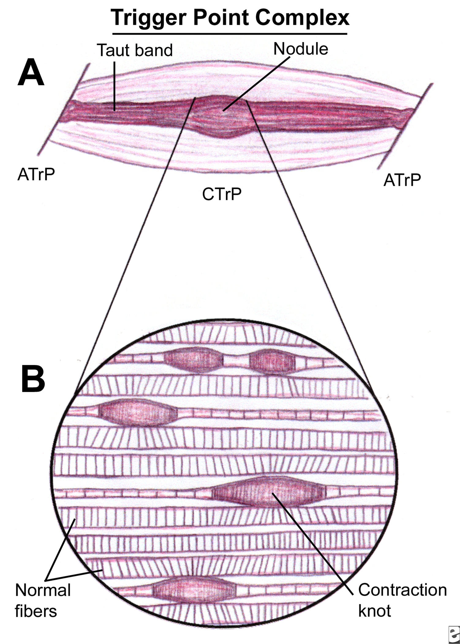
Trigger Points

What is a trigger point (TrP)?

Definition 1.

A focal hyperirritable spot in skeletal muscle associated with a palpable nodule in a taut band: painful on compression and can give rise to characteristic referred pain; tenderness; motor dysfunction and autonomic phenomena (Simons et al., 1999).

Definition 2.

"A trigger point is an area of the soft tissue which, after chronic contraction and reduced blood flow, becomes an area of high neurological activity. For example, fibers in an ischemic muscle (a muscle with low blood) can become an active trigger point in response to biochemical changes in the tissue. Active trigger points cause referred sensation to other parts of the body." www.painreliefvermont.com


Phases of Trigger Points (American Academy of Manual Medicine)

1. Active Trigger Point - Palpable nodular mass within muscles, fascia, scar tissue, ligaments, and periosteum. Produces a subjective referred pain complaint or autonomic phenomena, which is present constantly regardless of activity or rest.

2. (Same as above) produces a subjective referred pain or autonomic phenomena during activity but reduced or eliminated through rest.

3. Latent Trigger Point - (Same as above) but when snapped transversely it may replicate a referral pattern as if it were active. Without direct pressure, it does not produce a subjective referred pain or autonomic phenomena during either activity or rest.

4. Muscle tissue that does not exhibit a palpable ropy or button like nodular mass (characteristic of a TrP). However, the tissue may be hypertonic effecting  ROM, structural deviation, and hypersensitive to tactile pressure.   


Aetiological factors (what can cause them?)
Travell and Simons (1999)

Direct stimuli = Maggie Simpson Resists Talking

  • Mechanical overload
  • Sudden cooling of fatigued muscles
  • Repetitive usage resulting in fatigue
  • Trauma
Indirect stimuli = My Other VAN MET IN EGYPT
  • Myopathy
  • Other TrPs which activate secondary triggers
  • Visceral Disease
  • Arthropathy
  • Neuropathy
  • Metabolic dysfunction
  • Endocrine dysfunction
  • Toxicity
  • Infection
  • Emotional Distress

How can I treat them?

Good thing about trigger points is that they're predominantly self-treatable and also treatable with massage.

Lacrosse ball! (Check out this clip with Kelly Starrett going through some lower limb demonstrations of how to use the lacrosse ball for self-myofascial release)


Ischaemic compression (While the thumbnail of this vid looks dodgey as hell the explanation of this technique is useful in getting a better idea of how it fits in with what's going on at tissue level. There is no explanation about why he has his shirt off as well?!)


To make up for that last video, here's a chiropractor talking about locating pec minor/major trigger points.


These are just a couple of techniques that can be used. Pettrisage, deep stroking, vibrations are other ways of resolving trigger points but essentially the desired effect is the same, regardless of what technique you use: facilitate localised lymphatic drainage and enhance the flow of oxygenated blood (and therefore nutrients) to the ischaemic area of tissue. Also, as Kstar mentions the video above, we want to improve the ability of the muscle, fascia, nerves etc to slide along the mechanical interfaces of each other to allow for smooth full ROM at each joint.

To finish, Claire Davies uses an analogy that I really find helpful comparing trigger point release to wringing out a wet cloth. A repetetive 'milking' action to rinse out the dirt until fresh water can run through clear (Davies, 2002).


Davies, C (2002) Massage & Bodywork Magazine, Associate Bodywork and Massage Professionals.

Simons, D., Travell, J., Simons, L (1999) Myofascial Pain and Dysfunction: Upper half of body, Lippincott Williams & Wilkins.

Sunday, 1 January 2012

Isokinetic vs Isotonic

ISOKINETIC

This is when the muscle contracts at a constant speed and max effort through the full ROM. This can be achieved by a therapist when either assessing or treating by applying an active resistance to the patient/client but allowing them to overcome this at a certain speed through the entire range. A KIN-COM machine achieves the same effect, permitting the muscle to contract whilst keeping the same max force/rate movement through range (therefore building strength through range).

ISOTONIC

workThe force/tone remains constant BUT speed can vary.  This speed can vary based on lever length.
Disadvantage is isotonic training is that because there's that potential for variability muscles can develop predominantly in only the area where the most force is applied.


 http://ffden-2.phys.uaf.edu/211_fall2004.web.dir/kelsey_alexander/levers.html

Saturday, 31 December 2011

Cold/Cryotherapy/Constrast bathing


COLD/CRYOTHERAPY

The immediate aims of an acute soft tissue injury are to: Reduce pain/metabolic demand of tissues, limit/reduce inflammatory exudate, promote new tissue growth, protect newly forming tissue from disruption, maintain general levels of musculoskeletal/cardiorespiratory health (e.g. cycling/swimming). Cold therapy would come under the 'I' in P.R.I.C.E.D (protect, rest, ice, compression, elevation and the optional 'D' for drugs as in NSAIDS).

Application guidelines

10-20 minutes
Repeated every 2 waking hours over the acute/sub-acute stages

NB: This is dependant on the area injured (depth of tissue and how vascular it is). Ice can be used in any stage of the recovery but it's during this period that you'd generally apply it.

Contraindications

- Raynaud's disease
- Severe diabetes
- Cardiac problems
- Circulatory problems
- Elderly patients
- Radio/chemotherapy
- Hypersensitivity (hyperesthesia)

Beneficial physiological effects:
  1. Vasoconstriction reduces excessive bleeding into the site of injury site and therefore swelling. Excessive accumulation of swelling (oedema) can cause secondary hypoxic injury.  
  2. Reduces pain (via non-noxious adelta synaptic inhibition)
  3. Reduces muscle spasm
  4. Lessens risk of cell death by reducing metabolic rate

CONTRAST BATHING

Application guidelines

To be used during the sub-acute/chronic stages of soft tissue injury (NOT acute)

Showers: 1 - 2 mins hot followed by 1 - 30 secs cold (x 3 repetitions)
Baths: 3-4 mins hot followed by 30-60 secs cold (x 3 repetitions)

Early sub-acute: Begin with cold 3-4 mins then 1 min hot (repeat x 3 finishing on cold)
Durations of each phase can be altered depending on stage of healing (e.g. early or late sub-acute) but the basic aim is to reduce the cold and increase the hot (finishing on cold).

"As hot as you can bear and as cold as you can make it!"

How does it work?

The alternating of hot and cold temperatures aids the healing process by stimulating vasoconstriction/dilation which causes an increased peristaltic action (smooth muscle pump) flushing out waste products and aiding the transportation of nutrients and oxygen to the area. The changes in temperature have to be dramatic enough to achieve this effect i.e. cold in the range of 12-15 degrees and hot between 37-43 degrees.

Monday, 19 December 2011

Thursday, 8 December 2011

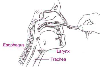
Tracheo-bronchial suction


Suction is used to 'suck up' the 'gunk' (secretions) sitting on your bronchial tree for patients that can't clear their own airway for themselves.


You can suction via...
  • Trachy (tracheostomy)
  • Endotracheal tube
  • Oral airway
  • Nasal airway (e.g. Nasopharangeal)

Indications (when to think about using it!)
  •  Reduced cough effort or inability to cough to clear airway
Contradications (when to NOT use it!)
  •  Frank haemoptysis (expectoration of blood only)
  • Severe bronchospasm
  • Undrained pneumothorax
  • Compromised CVS
  • Raised ICP
  • Fractured skull/facial bones
Hazards of Suctioning
  • Mucosal trauma
Suction is an invasive procedure and causes irritation to the delicate lining of the airways (tracheal/bronchial epithelium). This adverse effect is minimised by using an appropriate size catheter (simple calucation to figure this out: double size of trachy and minus 2 e.g. size 8 trachy = 16 - 2 = size 14 catheter). Also make sure to use correct pressures e.g. around 25.

  • Hypoxia
If ventilation is interupted (e.g. too big a tube, suctioning for too long etc) a reflex bronchospasm may occur. Pre and post Rx O2 can help reduce any hypoxia.

  • Cardiac arrthymias/bradycardia
Suctioning can cause a vasovagal reflex (a GI tract reflex that controls contraction of gastrointestinal muscles in response to stretching of the tract by food, controlled by the stretch/osmo & chemoreceptors of the dorsal vagal complex). This can lead to arrthymias so check with medics first.

  • Raised ICP
Suction raises ICP so if someone already has elevated levels (e.g. TBI, SAH etc) then be very cautious and seek medical opinion prior to attempting suction.


Thursday, 1 December 2011

FEV1/FVC ratio

This is a ratio used to diagnose OBSTRUCTIVE and RESTRICTIVE lung disease.

Obstructive pattern

In an obstructive lung disease like COPD most of the air can squeeze its way out eventually but takes a long time to do so (due to an obstruction!) This might be a sputum plug or floppy airways seen in conditions such as emphysema or tracheomalacia.

  • Problems with airflow into and out of the lungs
  • Increased RV (residual volume of air left in the lungs after expiration)
  • This air-trapping can lead to hyperinflation (barrell chest etc)

Effect of FEV1/FVC
  • Reduced FEV 1
  • FEV1/FVC ratio less than 70% of predicted value (ability to exhale 70% of breath in 1 sec)

Obstructive pattern


Due to conditions in which the airways are obstructed eg asthma or COPD. The FEV1 and FVC are reduced disproportionately.


Restrictive pattern

In an restrictive lung condition getting the air out isnt an issue, its the fact that there's not a whole lot of air to get out in the first place! If you imagine stiff hardened lungs that have difficulty expanding, you're not going to be able to inhale a lot of air to expel. Pathologies like pulmonary fibrosis, sarcoidosis, and certain types of pneumonia can have this effect.

  • Lung expansion/compliance is RESTRICTED...
  • Which causes decreased  lung volumes...
  • Which causes increase WOB...
  • Leading to reduced ventilation (reduced V of the V/Q ratio)
  • Annnd if breathing is harder work, patient is more likely to fatigue and start retaining CO2 which is never good.

Effect of FEV1/FVC
  • Reduced FEV1 and FVC equally

Restrictive pattern


Due to conditions in which the lung volume is reduced eg fibrosing alveolitis, scoliosis. The FEV1 and FVC are reduced proportionately.


National guidelines vary:

FEV1 as a % of predicted

  • <80% = mild
  • <60% = moderate
  • <40% = severe


Diagrams referenced from:
  • <80% = mild
  • <50% = moderate
  • <30% = severe
    (NICE guidelines)


http://www.gp-training.net/protocol/respiratory/copd/spirometry.htm

Functional overlay

"An emotional aspect of an organic disease. It may occur as an overreaction to an illness and is characterized by symptoms that continue long after clinical signs of the disease have ended."

Thursday, 24 November 2011

Spasticity vs rigidity vs hypertonicity


Hypertonicity: increased resistance to passive lengthening of a muscle. The underlying reason for the hypertonicity is not part of the definition. Hypertonicity could be due to a neural drive problem such as spasticity or rigidity or it could be due to increased resistance due to non contractile element changes in the musculotendinous unit such as contracture.

Spasticity is velocity dependent increased resistance to passive lengthening of the muscle: The faster you stretch the muscle the greater the resistance. Spasticity is clearly neural in nature and is  associated with the upper motor syndrome (UMN). Involvement of the corticospinal tract is often associated with the upper motor unit syndrome and spasticity. There are a number of clinical features that are also associated with spasticity that are part of the upper motor syndrome and some authors consider these features part of spasticity: associated reactions, hypereflexia, flexor synergy in the upper limb and extensor synergy in the lower limb. However including these phenomena as part of spasticity muddies the water so it is better to limit spasticity to velocity dependent resistance.

Rigidity
is increased neural activity throughout the range of muscle excursion and is not velocity dependent: present in both agonist and antagonist. This neural impairment is often associated with basal ganglia disease such as Parkinson’s disease. Lead pipe rigidity describes a constant resistance where when moving a joint resistance is felt and maintains the joint in the altered position. In cogwheel rigidity one feels the resistance rhythmically vary when applying a passive movement. It is thought to be the product of an underlying resting tremor which is masked by the rigidity but can be felt on passive movement.

In TBI you can get all sorts of combinations of neural impairments including spasticity and rigidity. Spasticity in TBI can also be very severe and may well be a somewhat different phenomena to what one sees in conditions like stroke or MS.

NB: Many thanks to gcoe from the physio forum for these definitions!

Saturday, 19 November 2011

The Pinky and the BRAIN


FRONTAL LOBE "Emotional control centre & home to our personality"
  • Motor function/initiation
  • Problem solving/judgement/decision making/reasoning
  • Social and sexual behaviour/impulse control
  • Spontaneity
  • Memory
  • Language


PARIETAL LOBE "What we feel & how we perceive"

1st functional area: Sensation and Perception
  • Integrates sensory information to form a single perception
2nd functional area: Spatial coordinate system 
  • Represents world around us
3rd Somatosensory cortex:
  • Processing of nerve impulses related to: touch, pain, temp, taste, pressure etc (information from mechanoreceptors, chemoreceptors, nociceptors)
TEMPORAL LOBE 'Primary Organisation of Sensory Input'
  • Hearing, Memory, Meaning, Language
  • Interpreting and processing auditory stimuli
  • Play a role in emotion and learning
  • Recognition of words
  • Memory of verbal material
OCCIPITAL LOBE 'Eyes in the back of our head'
  • Brain's ability to recognise objects
  • Vision

www.neuroskills.com
http://library.thinkquest.org

    Saturday, 12 November 2011

    Fish Oil (Omega 3) What's the deal?


    One of the most widely talked about supplements by fitness geeks and couch potatoes alike is fish oils. The ratio between omega 6 and omega 3 is out of balance with most in the Western world, said to be between 15-16.7 : 1  (Simopoulos, 2002) when ideally it should be more like 2:1 (the better the ratio the better the health benefits!)

    EFAs are 'essential fatty acids' named as such because we can't produce them in our bodies meaning we need to include them in diet. So we do need both omega 6 and 3 but you've probably got enough 6 from what you already eat (safflower oil, sunflower oil, corn oil) these guys will regularly crop up on the ingredients list of processed foods and are not limited to just plant sources! If you're eating beef from a cow that's grain fed, you got yourself some omega 6 right there (as opposed to the ideal grass fed animals who are higher in omega 3).

    EPA - Eicosapentaenoic acid
    DHA - Docosahexaenoic acid

    These are essential fatty acid components of omega 3 that provide some of the many health benefits derived from fish oil intake. These include:

    1) Improved insulin sensitivity in muscle cells / Decreased insulin sensitivity in fat cells - suggesting that nutrient intake from food would be inclined to protein storage rather than being stored in adipose tissue. This would potentially increase metabolic rate and lean mass (Berardi and Mejia, 2005).

    2) Reduced risk of cardiovascular disease, cancer, diabetes (Simopoulos, 2002)

    3) Improved fetal brain development - the addition of DHA to a pregnant woman's diet may be beneficial for the fetal brain development (Hoffman, 2011)

    EPA and DHA are important components of retina and brain tissue and can help to form neuro-transmitters

    The health benefits are not just limited to the diseases above. Simopoulos (2002) showed in a study that the improved omega 6 to 3 ratio could have beneficial effects on other pathologies. A ratio of 2.5:1 reduced rectal cell proliferation in patients with colorectal cancer compared to a ratio of 4:1 that did not prove to have the same effect. In inflammatory diseases such as rheumatoid arthritis a ratio of 2-3:1 supressed inflammation. A ratio of 5:1 had positive implications on asthma sufferers whereas a ratio of 10:1 showed adverse effects.

    A 'perfect ratio' does not therefore stand out as the diseases listed above are multifactorial and cannot all be improved a universal ratio it would seem. However, the general consensus seems to be that a lower and more balanced ratio of omega 6 to 3 has positive health outcomes whereas a higher omega 6 to 3 can have adverse effects on the body. What's more is taking fish oil as a supplement has no health risks and will only serve to benefit your health (Berardi and Mejia, 2005). So it's a no-brainer guys, if you're not on this stuff already, start taking it or at least start eating more oily fish!


    Does flaxseed have the same benefits as taking fish oil?  

    Apparently not. There are plant sources of omega 3s such as flax, hempseed and walnut oil contain alpha-linoleic acid*. Your body can convert this into EPA and DHA but there is said to be very limited metabolic conversion of dietary alpha-linoleic acid to DHA. These plant sources themselves have no DHA content (Omega 3 institute, 2010).

    *Please note that alpha linoleic acid is not the same thing as alpha lipoic acid which is an antioxidant produced in the body that aids glucose conversion into energy.

    Simopoulos, AP (2002) 'The importance of the ratio of omega-6/omega-3 essential fatty acids', Biomed Pharmacother, 56(8):365-79
    Berardi, J; Mejia, M (2005) 'Scrawny to Brawny' , Rodale, USA