5 = Normal (100%) Complete ROM against gravity with maximal resistance
4 = Good (75%) Complete ROM against gravity with moderate resistance
3+ = Fair + Complete ROM against gravity with minimal resistance
3 = Fair (50%) Complete ROM against gravity
3 - = Fair - Some but not complete ROM against gravity
2 + = Poor + Initiates movement against gravity
2 = Poor Poor (25%) Complete ROM with gravity counter-balanced
2 - = Poor - Initiates motion IF gravity is counter-balanced
1 = Trace Flicker of contraction but not joint movement
0 = Nothing No contraction palpated
Tuesday, 24 April 2012

Wednesday, 4 January 2012
Trigger Points
What is a trigger point (TrP)?
Definition 1.
 A focal hyperirritable spot in skeletal muscle associated with a palpable nodule in a taut band: painful on compression and can give rise to characteristic referred pain; tenderness; motor dysfunction and autonomic phenomena (Simons et al., 1999).
Definition 2.
"A trigger point is an area of the soft tissue which, after chronic contraction and reduced blood flow, becomes an area of high neurological activity. For example, fibers in an ischemic muscle (a muscle with low blood) can become an active trigger point in response to biochemical changes in the tissue. Active trigger points cause referred sensation to other parts of the body." www.painreliefvermont.com
Phases of Trigger Points (American Academy of Manual Medicine)
1. Active Trigger Point - Palpable nodular mass within muscles, fascia, scar tissue, ligaments, and periosteum. Produces a subjective referred pain complaint or autonomic phenomena, which is present constantly regardless of activity or rest.
2. (Same as above) produces a subjective referred pain or autonomic phenomena during activity but reduced or eliminated through rest.
3. Latent Trigger Point - (Same as above) but when snapped transversely it may replicate a referral pattern as if it were active. Without direct pressure, it does not produce a subjective referred pain or autonomic phenomena during either activity or rest.
4. Muscle tissue that does not exhibit a palpable ropy or button like nodular mass (characteristic of a TrP). However, the tissue may be hypertonic effecting ROM, structural deviation, and hypersensitive to tactile pressure.

Aetiological factors (what can cause them?)
Travell and Simons (1999)
Direct stimuli = Maggie Simpson Resists Talking
How can I treat them?
Good thing about trigger points is that they're predominantly self-treatable and also treatable with massage.
Lacrosse ball! (Check out this clip with Kelly Starrett going through some lower limb demonstrations of how to use the lacrosse ball for self-myofascial release)
Ischaemic compression (While the thumbnail of this vid looks dodgey as hell the explanation of this technique is useful in getting a better idea of how it fits in with what's going on at tissue level. There is no explanation about why he has his shirt off as well?!)

Definition 1.
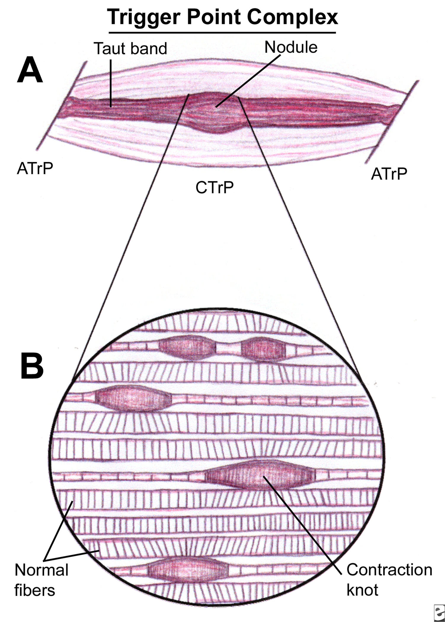 A focal hyperirritable spot in skeletal muscle associated with a palpable nodule in a taut band: painful on compression and can give rise to characteristic referred pain; tenderness; motor dysfunction and autonomic phenomena (Simons et al., 1999).
 Definition 2.
"A trigger point is an area of the soft tissue which, after chronic contraction and reduced blood flow, becomes an area of high neurological activity. For example, fibers in an ischemic muscle (a muscle with low blood) can become an active trigger point in response to biochemical changes in the tissue. Active trigger points cause referred sensation to other parts of the body." www.painreliefvermont.com
Phases of Trigger Points (American Academy of Manual Medicine)
1. Active Trigger Point - Palpable nodular mass within muscles, fascia, scar tissue, ligaments, and periosteum. Produces a subjective referred pain complaint or autonomic phenomena, which is present constantly regardless of activity or rest.
2. (Same as above) produces a subjective referred pain or autonomic phenomena during activity but reduced or eliminated through rest.
3. Latent Trigger Point - (Same as above) but when snapped transversely it may replicate a referral pattern as if it were active. Without direct pressure, it does not produce a subjective referred pain or autonomic phenomena during either activity or rest.
4. Muscle tissue that does not exhibit a palpable ropy or button like nodular mass (characteristic of a TrP). However, the tissue may be hypertonic effecting ROM, structural deviation, and hypersensitive to tactile pressure.
Aetiological factors (what can cause them?)
Travell and Simons (1999)
Direct stimuli = Maggie Simpson Resists Talking
- Mechanical overload
- Sudden cooling of fatigued muscles
- Repetitive usage resulting in fatigue
- Trauma
- Myopathy
- Other TrPs which activate secondary triggers
- Visceral Disease
- Arthropathy
- Neuropathy
- Metabolic dysfunction
- Endocrine dysfunction
- Toxicity
- Infection
- Emotional Distress

How can I treat them?
Good thing about trigger points is that they're predominantly self-treatable and also treatable with massage.
Lacrosse ball! (Check out this clip with Kelly Starrett going through some lower limb demonstrations of how to use the lacrosse ball for self-myofascial release)
Ischaemic compression (While the thumbnail of this vid looks dodgey as hell the explanation of this technique is useful in getting a better idea of how it fits in with what's going on at tissue level. There is no explanation about why he has his shirt off as well?!)
To make up for that last video, here's a chiropractor talking about locating pec minor/major trigger points.
These are just a couple of techniques that can be used. Pettrisage, deep stroking, vibrations are other ways of resolving trigger points but essentially the desired effect is the same, regardless of what technique you use: facilitate localised lymphatic drainage and enhance the flow of oxygenated blood (and therefore nutrients) to the ischaemic area of tissue. Also, as Kstar mentions the video above, we want to improve the ability of the muscle, fascia, nerves etc to slide along the mechanical interfaces of each other to allow for smooth full ROM at each joint.
To finish, Claire Davies uses an analogy that I really find helpful comparing trigger point release to wringing out a wet cloth. A repetetive 'milking' action to rinse out the dirt until fresh water can run through clear (Davies, 2002).
Davies, C (2002) Massage & Bodywork Magazine, Associate Bodywork and Massage Professionals.
Simons, D., Travell, J., Simons, L (1999) Myofascial Pain and Dysfunction: Upper half of body, Lippincott Williams & Wilkins.
Sunday, 1 January 2012
Isokinetic vs Isotonic
ISOKINETIC
This is when the muscle contracts at a constant speed and max effort through the full ROM. This can be achieved by a therapist when either assessing or treating by applying an active resistance to the patient/client but allowing them to overcome this at a certain speed through the entire range. A KIN-COM machine achieves the same effect, permitting the muscle to contract whilst keeping the same max force/rate movement through range (therefore building strength through range).
ISOTONIC
 The force/tone remains constant BUT speed can vary. This speed can vary based on lever length.
Disadvantage is isotonic training is that because there's that potential for variability muscles can develop predominantly in only the area where the most force is applied.
http://ffden-2.phys.uaf.edu/211_fall2004.web.dir/kelsey_alexander/levers.html
This is when the muscle contracts at a constant speed and max effort through the full ROM. This can be achieved by a therapist when either assessing or treating by applying an active resistance to the patient/client but allowing them to overcome this at a certain speed through the entire range. A KIN-COM machine achieves the same effect, permitting the muscle to contract whilst keeping the same max force/rate movement through range (therefore building strength through range).
ISOTONIC
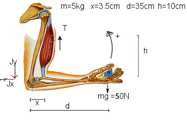 The force/tone remains constant BUT speed can vary. This speed can vary based on lever length.
 Disadvantage is isotonic training is that because there's that potential for variability muscles can develop predominantly in only the area where the most force is applied.
http://ffden-2.phys.uaf.edu/211_fall2004.web.dir/kelsey_alexander/levers.html
Saturday, 31 December 2011
Cold/Cryotherapy/Constrast bathing
 COLD/CRYOTHERAPY
The immediate aims of an acute soft tissue injury are to: Reduce pain/metabolic demand of tissues, limit/reduce inflammatory exudate, promote new tissue growth, protect newly forming tissue from disruption, maintain general levels of musculoskeletal/cardiorespiratory health (e.g. cycling/swimming). Cold therapy would come under the 'I' in P.R.I.C.E.D (protect, rest, ice, compression, elevation and the optional 'D' for drugs as in NSAIDS).
Application guidelines
10-20 minutes
Repeated every 2 waking hours over the acute/sub-acute stages
NB: This is dependant on the area injured (depth of tissue and how vascular it is). Ice can be used in any stage of the recovery but it's during this period that you'd generally apply it.
Contraindications
- Raynaud's disease
- Severe diabetes
- Cardiac problems
- Circulatory problems
- Elderly patients
- Radio/chemotherapy
- Hypersensitivity (hyperesthesia)
Beneficial physiological effects:
- Vasoconstriction reduces excessive bleeding into the site of injury site and therefore swelling. Excessive accumulation of swelling (oedema) can cause secondary hypoxic injury.
- Reduces pain (via non-noxious adelta synaptic inhibition)
- Reduces muscle spasm
- Lessens risk of cell death by reducing metabolic rate
CONTRAST BATHING
Application guidelines
To be used during the sub-acute/chronic stages of soft tissue injury (NOT acute)
 Showers: 1 - 2 mins hot followed by 1 - 30 secs cold (x 3 repetitions)
 Baths: 3-4 mins hot followed by 30-60 secs cold (x 3 repetitions)
Early sub-acute: Begin with cold 3-4 mins then 1 min hot (repeat x 3 finishing on cold)
Durations of each phase can be altered depending on stage of healing (e.g. early or late sub-acute) but the basic aim is to reduce the cold and increase the hot (finishing on cold).
"As hot as you can bear and as cold as you can make it!"
How does it work?
The alternating of hot and cold temperatures aids the healing process by stimulating vasoconstriction/dilation which causes an increased peristaltic action (smooth muscle pump) flushing out waste products and aiding the transportation of nutrients and oxygen to the area. The changes in temperature have to be dramatic enough to achieve this effect i.e. cold in the range of 12-15 degrees and hot between 37-43 degrees.
Monday, 19 December 2011
Thursday, 8 December 2011
Tracheo-bronchial suction

Suction is used to 'suck up' the 'gunk' (secretions) sitting on your bronchial tree for patients that can't clear their own airway for themselves.
You can suction via...
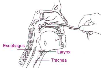
- Trachy (tracheostomy)
- Endotracheal tube
- Oral airway
- Nasal airway (e.g. Nasopharangeal)
Indications (when to think about using it!)
- Reduced cough effort or inability to cough to clear airway
Contradications (when to NOT use it!)
- Frank haemoptysis (expectoration of blood only)
- Severe bronchospasm
- Undrained pneumothorax
- Compromised CVS
- Raised ICP
- Fractured skull/facial bones
- Mucosal trauma
- Hypoxia
- Cardiac arrthymias/bradycardia
- Raised ICP
Thursday, 1 December 2011
FEV1/FVC ratio
This is a ratio used to diagnose OBSTRUCTIVE and RESTRICTIVE lung disease.
Obstructive pattern
In an obstructive lung disease like COPD most of the air can squeeze its way out eventually but takes a long time to do so (due to an obstruction!) This might be a sputum plug or floppy airways seen in conditions such as emphysema or tracheomalacia.
Effect of FEV1/FVC
Due to conditions in which the airways are obstructed eg asthma or COPD. The FEV1 and FVC are reduced disproportionately.

Restrictive pattern
In an restrictive lung condition getting the air out isnt an issue, its the fact that there's not a whole lot of air to get out in the first place! If you imagine stiff hardened lungs that have difficulty expanding, you're not going to be able to inhale a lot of air to expel. Pathologies like pulmonary fibrosis, sarcoidosis, and certain types of pneumonia can have this effect.
Effect of FEV1/FVC
Due to conditions in which the lung volume is reduced eg fibrosing alveolitis, scoliosis. The FEV1 and FVC are reduced proportionately.

National guidelines vary:
FEV1 as a % of predicted
Obstructive pattern
In an obstructive lung disease like COPD most of the air can squeeze its way out eventually but takes a long time to do so (due to an obstruction!) This might be a sputum plug or floppy airways seen in conditions such as emphysema or tracheomalacia.
- Problems with airflow into and out of the lungs
- Increased RV (residual volume of air left in the lungs after expiration)
- This air-trapping can lead to hyperinflation (barrell chest etc)
Effect of FEV1/FVC
- Reduced FEV 1
- FEV1/FVC ratio less than 70% of predicted value (ability to exhale 70% of breath in 1 sec)
Obstructive pattern
Due to conditions in which the airways are obstructed eg asthma or COPD. The FEV1 and FVC are reduced disproportionately.
Restrictive pattern
In an restrictive lung condition getting the air out isnt an issue, its the fact that there's not a whole lot of air to get out in the first place! If you imagine stiff hardened lungs that have difficulty expanding, you're not going to be able to inhale a lot of air to expel. Pathologies like pulmonary fibrosis, sarcoidosis, and certain types of pneumonia can have this effect.
- Lung expansion/compliance is RESTRICTED...
- Which causes decreased lung volumes...
- Which causes increase WOB...
- Leading to reduced ventilation (reduced V of the V/Q ratio)
- Annnd if breathing is harder work, patient is more likely to fatigue and start retaining CO2 which is never good.
Effect of FEV1/FVC
- Reduced FEV1 and FVC equally
Restrictive pattern
Due to conditions in which the lung volume is reduced eg fibrosing alveolitis, scoliosis. The FEV1 and FVC are reduced proportionately.
National guidelines vary:
FEV1 as a % of predicted
Diagrams referenced from: |
http://www.gp-training.net/protocol/respiratory/copd/spirometry.htm |
Subscribe to:
Posts (Atom)