Saturday 31 December 2011

Cold/Cryotherapy/Constrast bathing


COLD/CRYOTHERAPY

The immediate aims of an acute soft tissue injury are to: Reduce pain/metabolic demand of tissues, limit/reduce inflammatory exudate, promote new tissue growth, protect newly forming tissue from disruption, maintain general levels of musculoskeletal/cardiorespiratory health (e.g. cycling/swimming). Cold therapy would come under the 'I' in P.R.I.C.E.D (protect, rest, ice, compression, elevation and the optional 'D' for drugs as in NSAIDS).

Application guidelines

10-20 minutes
Repeated every 2 waking hours over the acute/sub-acute stages

NB: This is dependant on the area injured (depth of tissue and how vascular it is). Ice can be used in any stage of the recovery but it's during this period that you'd generally apply it.

Contraindications

- Raynaud's disease
- Severe diabetes
- Cardiac problems
- Circulatory problems
- Elderly patients
- Radio/chemotherapy
- Hypersensitivity (hyperesthesia)

Beneficial physiological effects:
  1. Vasoconstriction reduces excessive bleeding into the site of injury site and therefore swelling. Excessive accumulation of swelling (oedema) can cause secondary hypoxic injury.  
  2. Reduces pain (via non-noxious adelta synaptic inhibition)
  3. Reduces muscle spasm
  4. Lessens risk of cell death by reducing metabolic rate

CONTRAST BATHING

Application guidelines

To be used during the sub-acute/chronic stages of soft tissue injury (NOT acute)

Showers: 1 - 2 mins hot followed by 1 - 30 secs cold (x 3 repetitions)
Baths: 3-4 mins hot followed by 30-60 secs cold (x 3 repetitions)

Early sub-acute: Begin with cold 3-4 mins then 1 min hot (repeat x 3 finishing on cold)
Durations of each phase can be altered depending on stage of healing (e.g. early or late sub-acute) but the basic aim is to reduce the cold and increase the hot (finishing on cold).

"As hot as you can bear and as cold as you can make it!"

How does it work?

The alternating of hot and cold temperatures aids the healing process by stimulating vasoconstriction/dilation which causes an increased peristaltic action (smooth muscle pump) flushing out waste products and aiding the transportation of nutrients and oxygen to the area. The changes in temperature have to be dramatic enough to achieve this effect i.e. cold in the range of 12-15 degrees and hot between 37-43 degrees.

Monday 19 December 2011

Thursday 8 December 2011

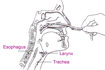
Tracheo-bronchial suction


Suction is used to 'suck up' the 'gunk' (secretions) sitting on your bronchial tree for patients that can't clear their own airway for themselves.


You can suction via...
  • Trachy (tracheostomy)
  • Endotracheal tube
  • Oral airway
  • Nasal airway (e.g. Nasopharangeal)

Indications (when to think about using it!)
  •  Reduced cough effort or inability to cough to clear airway
Contradications (when to NOT use it!)
  •  Frank haemoptysis (expectoration of blood only)
  • Severe bronchospasm
  • Undrained pneumothorax
  • Compromised CVS
  • Raised ICP
  • Fractured skull/facial bones
Hazards of Suctioning
  • Mucosal trauma
Suction is an invasive procedure and causes irritation to the delicate lining of the airways (tracheal/bronchial epithelium). This adverse effect is minimised by using an appropriate size catheter (simple calucation to figure this out: double size of trachy and minus 2 e.g. size 8 trachy = 16 - 2 = size 14 catheter). Also make sure to use correct pressures e.g. around 25.

  • Hypoxia
If ventilation is interupted (e.g. too big a tube, suctioning for too long etc) a reflex bronchospasm may occur. Pre and post Rx O2 can help reduce any hypoxia.

  • Cardiac arrthymias/bradycardia
Suctioning can cause a vasovagal reflex (a GI tract reflex that controls contraction of gastrointestinal muscles in response to stretching of the tract by food, controlled by the stretch/osmo & chemoreceptors of the dorsal vagal complex). This can lead to arrthymias so check with medics first.

  • Raised ICP
Suction raises ICP so if someone already has elevated levels (e.g. TBI, SAH etc) then be very cautious and seek medical opinion prior to attempting suction.


Thursday 1 December 2011

FEV1/FVC ratio

This is a ratio used to diagnose OBSTRUCTIVE and RESTRICTIVE lung disease.

Obstructive pattern

In an obstructive lung disease like COPD most of the air can squeeze its way out eventually but takes a long time to do so (due to an obstruction!) This might be a sputum plug or floppy airways seen in conditions such as emphysema or tracheomalacia.

  • Problems with airflow into and out of the lungs
  • Increased RV (residual volume of air left in the lungs after expiration)
  • This air-trapping can lead to hyperinflation (barrell chest etc)

Effect of FEV1/FVC
  • Reduced FEV 1
  • FEV1/FVC ratio less than 70% of predicted value (ability to exhale 70% of breath in 1 sec)

Obstructive pattern


Due to conditions in which the airways are obstructed eg asthma or COPD. The FEV1 and FVC are reduced disproportionately.


Restrictive pattern

In an restrictive lung condition getting the air out isnt an issue, its the fact that there's not a whole lot of air to get out in the first place! If you imagine stiff hardened lungs that have difficulty expanding, you're not going to be able to inhale a lot of air to expel. Pathologies like pulmonary fibrosis, sarcoidosis, and certain types of pneumonia can have this effect.

  • Lung expansion/compliance is RESTRICTED...
  • Which causes decreased  lung volumes...
  • Which causes increase WOB...
  • Leading to reduced ventilation (reduced V of the V/Q ratio)
  • Annnd if breathing is harder work, patient is more likely to fatigue and start retaining CO2 which is never good.

Effect of FEV1/FVC
  • Reduced FEV1 and FVC equally

Restrictive pattern


Due to conditions in which the lung volume is reduced eg fibrosing alveolitis, scoliosis. The FEV1 and FVC are reduced proportionately.


National guidelines vary:

FEV1 as a % of predicted

  • <80% = mild
  • <60% = moderate
  • <40% = severe


Diagrams referenced from:
  • <80% = mild
  • <50% = moderate
  • <30% = severe
    (NICE guidelines)


http://www.gp-training.net/protocol/respiratory/copd/spirometry.htm

Functional overlay

"An emotional aspect of an organic disease. It may occur as an overreaction to an illness and is characterized by symptoms that continue long after clinical signs of the disease have ended."