Saturday, 31 December 2011

Cold/Cryotherapy/Constrast bathing


COLD/CRYOTHERAPY

The immediate aims of an acute soft tissue injury are to: Reduce pain/metabolic demand of tissues, limit/reduce inflammatory exudate, promote new tissue growth, protect newly forming tissue from disruption, maintain general levels of musculoskeletal/cardiorespiratory health (e.g. cycling/swimming). Cold therapy would come under the 'I' in P.R.I.C.E.D (protect, rest, ice, compression, elevation and the optional 'D' for drugs as in NSAIDS).

Application guidelines

10-20 minutes
Repeated every 2 waking hours over the acute/sub-acute stages

NB: This is dependant on the area injured (depth of tissue and how vascular it is). Ice can be used in any stage of the recovery but it's during this period that you'd generally apply it.

Contraindications

- Raynaud's disease
- Severe diabetes
- Cardiac problems
- Circulatory problems
- Elderly patients
- Radio/chemotherapy
- Hypersensitivity (hyperesthesia)

Beneficial physiological effects:
  1. Vasoconstriction reduces excessive bleeding into the site of injury site and therefore swelling. Excessive accumulation of swelling (oedema) can cause secondary hypoxic injury.  
  2. Reduces pain (via non-noxious adelta synaptic inhibition)
  3. Reduces muscle spasm
  4. Lessens risk of cell death by reducing metabolic rate

CONTRAST BATHING

Application guidelines

To be used during the sub-acute/chronic stages of soft tissue injury (NOT acute)

Showers: 1 - 2 mins hot followed by 1 - 30 secs cold (x 3 repetitions)
Baths: 3-4 mins hot followed by 30-60 secs cold (x 3 repetitions)

Early sub-acute: Begin with cold 3-4 mins then 1 min hot (repeat x 3 finishing on cold)
Durations of each phase can be altered depending on stage of healing (e.g. early or late sub-acute) but the basic aim is to reduce the cold and increase the hot (finishing on cold).

"As hot as you can bear and as cold as you can make it!"

How does it work?

The alternating of hot and cold temperatures aids the healing process by stimulating vasoconstriction/dilation which causes an increased peristaltic action (smooth muscle pump) flushing out waste products and aiding the transportation of nutrients and oxygen to the area. The changes in temperature have to be dramatic enough to achieve this effect i.e. cold in the range of 12-15 degrees and hot between 37-43 degrees.

Monday, 19 December 2011

Thursday, 8 December 2011

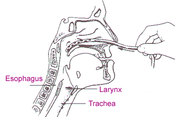
Tracheo-bronchial suction


Suction is used to 'suck up' the 'gunk' (secretions) sitting on your bronchial tree for patients that can't clear their own airway for themselves.


You can suction via...
  • Trachy (tracheostomy)
  • Endotracheal tube
  • Oral airway
  • Nasal airway (e.g. Nasopharangeal)

Indications (when to think about using it!)
  •  Reduced cough effort or inability to cough to clear airway
Contradications (when to NOT use it!)
  •  Frank haemoptysis (expectoration of blood only)
  • Severe bronchospasm
  • Undrained pneumothorax
  • Compromised CVS
  • Raised ICP
  • Fractured skull/facial bones
Hazards of Suctioning
  • Mucosal trauma
Suction is an invasive procedure and causes irritation to the delicate lining of the airways (tracheal/bronchial epithelium). This adverse effect is minimised by using an appropriate size catheter (simple calucation to figure this out: double size of trachy and minus 2 e.g. size 8 trachy = 16 - 2 = size 14 catheter). Also make sure to use correct pressures e.g. around 25.

  • Hypoxia
If ventilation is interupted (e.g. too big a tube, suctioning for too long etc) a reflex bronchospasm may occur. Pre and post Rx O2 can help reduce any hypoxia.

  • Cardiac arrthymias/bradycardia
Suctioning can cause a vasovagal reflex (a GI tract reflex that controls contraction of gastrointestinal muscles in response to stretching of the tract by food, controlled by the stretch/osmo & chemoreceptors of the dorsal vagal complex). This can lead to arrthymias so check with medics first.

  • Raised ICP
Suction raises ICP so if someone already has elevated levels (e.g. TBI, SAH etc) then be very cautious and seek medical opinion prior to attempting suction.


Thursday, 1 December 2011

FEV1/FVC ratio

This is a ratio used to diagnose OBSTRUCTIVE and RESTRICTIVE lung disease.

Obstructive pattern

In an obstructive lung disease like COPD most of the air can squeeze its way out eventually but takes a long time to do so (due to an obstruction!) This might be a sputum plug or floppy airways seen in conditions such as emphysema or tracheomalacia.

  • Problems with airflow into and out of the lungs
  • Increased RV (residual volume of air left in the lungs after expiration)
  • This air-trapping can lead to hyperinflation (barrell chest etc)

Effect of FEV1/FVC
  • Reduced FEV 1
  • FEV1/FVC ratio less than 70% of predicted value (ability to exhale 70% of breath in 1 sec)

Obstructive pattern


Due to conditions in which the airways are obstructed eg asthma or COPD. The FEV1 and FVC are reduced disproportionately.


Restrictive pattern

In an restrictive lung condition getting the air out isnt an issue, its the fact that there's not a whole lot of air to get out in the first place! If you imagine stiff hardened lungs that have difficulty expanding, you're not going to be able to inhale a lot of air to expel. Pathologies like pulmonary fibrosis, sarcoidosis, and certain types of pneumonia can have this effect.

  • Lung expansion/compliance is RESTRICTED...
  • Which causes decreased  lung volumes...
  • Which causes increase WOB...
  • Leading to reduced ventilation (reduced V of the V/Q ratio)
  • Annnd if breathing is harder work, patient is more likely to fatigue and start retaining CO2 which is never good.

Effect of FEV1/FVC
  • Reduced FEV1 and FVC equally

Restrictive pattern


Due to conditions in which the lung volume is reduced eg fibrosing alveolitis, scoliosis. The FEV1 and FVC are reduced proportionately.


National guidelines vary:

FEV1 as a % of predicted

  • <80% = mild
  • <60% = moderate
  • <40% = severe


Diagrams referenced from:
  • <80% = mild
  • <50% = moderate
  • <30% = severe
    (NICE guidelines)


http://www.gp-training.net/protocol/respiratory/copd/spirometry.htm

Functional overlay

"An emotional aspect of an organic disease. It may occur as an overreaction to an illness and is characterized by symptoms that continue long after clinical signs of the disease have ended."

Thursday, 24 November 2011

Spasticity vs rigidity vs hypertonicity


Hypertonicity: increased resistance to passive lengthening of a muscle. The underlying reason for the hypertonicity is not part of the definition. Hypertonicity could be due to a neural drive problem such as spasticity or rigidity or it could be due to increased resistance due to non contractile element changes in the musculotendinous unit such as contracture.

Spasticity is velocity dependent increased resistance to passive lengthening of the muscle: The faster you stretch the muscle the greater the resistance. Spasticity is clearly neural in nature and is  associated with the upper motor syndrome (UMN). Involvement of the corticospinal tract is often associated with the upper motor unit syndrome and spasticity. There are a number of clinical features that are also associated with spasticity that are part of the upper motor syndrome and some authors consider these features part of spasticity: associated reactions, hypereflexia, flexor synergy in the upper limb and extensor synergy in the lower limb. However including these phenomena as part of spasticity muddies the water so it is better to limit spasticity to velocity dependent resistance.

Rigidity
is increased neural activity throughout the range of muscle excursion and is not velocity dependent: present in both agonist and antagonist. This neural impairment is often associated with basal ganglia disease such as Parkinson’s disease. Lead pipe rigidity describes a constant resistance where when moving a joint resistance is felt and maintains the joint in the altered position. In cogwheel rigidity one feels the resistance rhythmically vary when applying a passive movement. It is thought to be the product of an underlying resting tremor which is masked by the rigidity but can be felt on passive movement.

In TBI you can get all sorts of combinations of neural impairments including spasticity and rigidity. Spasticity in TBI can also be very severe and may well be a somewhat different phenomena to what one sees in conditions like stroke or MS.

NB: Many thanks to gcoe from the physio forum for these definitions!

Saturday, 19 November 2011

The Pinky and the BRAIN


FRONTAL LOBE "Emotional control centre & home to our personality"
  • Motor function/initiation
  • Problem solving/judgement/decision making/reasoning
  • Social and sexual behaviour/impulse control
  • Spontaneity
  • Memory
  • Language


PARIETAL LOBE "What we feel & how we perceive"

1st functional area: Sensation and Perception
  • Integrates sensory information to form a single perception
2nd functional area: Spatial coordinate system 
  • Represents world around us
3rd Somatosensory cortex:
  • Processing of nerve impulses related to: touch, pain, temp, taste, pressure etc (information from mechanoreceptors, chemoreceptors, nociceptors)
TEMPORAL LOBE 'Primary Organisation of Sensory Input'
  • Hearing, Memory, Meaning, Language
  • Interpreting and processing auditory stimuli
  • Play a role in emotion and learning
  • Recognition of words
  • Memory of verbal material
OCCIPITAL LOBE 'Eyes in the back of our head'
  • Brain's ability to recognise objects
  • Vision

www.neuroskills.com
http://library.thinkquest.org

    Saturday, 12 November 2011

    Fish Oil (Omega 3) What's the deal?


    One of the most widely talked about supplements by fitness geeks and couch potatoes alike is fish oils. The ratio between omega 6 and omega 3 is out of balance with most in the Western world, said to be between 15-16.7 : 1  (Simopoulos, 2002) when ideally it should be more like 2:1 (the better the ratio the better the health benefits!)

    EFAs are 'essential fatty acids' named as such because we can't produce them in our bodies meaning we need to include them in diet. So we do need both omega 6 and 3 but you've probably got enough 6 from what you already eat (safflower oil, sunflower oil, corn oil) these guys will regularly crop up on the ingredients list of processed foods and are not limited to just plant sources! If you're eating beef from a cow that's grain fed, you got yourself some omega 6 right there (as opposed to the ideal grass fed animals who are higher in omega 3).

    EPA - Eicosapentaenoic acid
    DHA - Docosahexaenoic acid

    These are essential fatty acid components of omega 3 that provide some of the many health benefits derived from fish oil intake. These include:

    1) Improved insulin sensitivity in muscle cells / Decreased insulin sensitivity in fat cells - suggesting that nutrient intake from food would be inclined to protein storage rather than being stored in adipose tissue. This would potentially increase metabolic rate and lean mass (Berardi and Mejia, 2005).

    2) Reduced risk of cardiovascular disease, cancer, diabetes (Simopoulos, 2002)

    3) Improved fetal brain development - the addition of DHA to a pregnant woman's diet may be beneficial for the fetal brain development (Hoffman, 2011)

    EPA and DHA are important components of retina and brain tissue and can help to form neuro-transmitters

    The health benefits are not just limited to the diseases above. Simopoulos (2002) showed in a study that the improved omega 6 to 3 ratio could have beneficial effects on other pathologies. A ratio of 2.5:1 reduced rectal cell proliferation in patients with colorectal cancer compared to a ratio of 4:1 that did not prove to have the same effect. In inflammatory diseases such as rheumatoid arthritis a ratio of 2-3:1 supressed inflammation. A ratio of 5:1 had positive implications on asthma sufferers whereas a ratio of 10:1 showed adverse effects.

    A 'perfect ratio' does not therefore stand out as the diseases listed above are multifactorial and cannot all be improved a universal ratio it would seem. However, the general consensus seems to be that a lower and more balanced ratio of omega 6 to 3 has positive health outcomes whereas a higher omega 6 to 3 can have adverse effects on the body. What's more is taking fish oil as a supplement has no health risks and will only serve to benefit your health (Berardi and Mejia, 2005). So it's a no-brainer guys, if you're not on this stuff already, start taking it or at least start eating more oily fish!


    Does flaxseed have the same benefits as taking fish oil?  

    Apparently not. There are plant sources of omega 3s such as flax, hempseed and walnut oil contain alpha-linoleic acid*. Your body can convert this into EPA and DHA but there is said to be very limited metabolic conversion of dietary alpha-linoleic acid to DHA. These plant sources themselves have no DHA content (Omega 3 institute, 2010).

    *Please note that alpha linoleic acid is not the same thing as alpha lipoic acid which is an antioxidant produced in the body that aids glucose conversion into energy.

    Simopoulos, AP (2002) 'The importance of the ratio of omega-6/omega-3 essential fatty acids', Biomed Pharmacother, 56(8):365-79
    Berardi, J; Mejia, M (2005) 'Scrawny to Brawny' , Rodale, USA

    Friday, 23 September 2011

    What the hell causes patellofemoral pain? The ominous VMOoooo!

    Sorry it's been so long since I last made a post. I tried making one just a second ago and when I finished it wouldn't publish and then it disappeared so this is my 2nd attempt at the same one! I've also just discovered the comments that some people have posted which was a nice surprise so thanks very much for that and if you'd like to see anything in particular then give me a shout!

    I've always been interested in finding out more about patellofemoral pain and just what exactly is the root cause of it. Is there just one? Can it be a combination of things? A lot of therapists/trainers are crazy about the VMO idea and a lot aren't so crazy. I had a patient whilst on placement who was complaining of patellofemoral symptoms. My educator encouraged me to focus on glute medius strengthening rather than VMO exercises to solve the problem. It's a common complaint among the general population and in athletes as they squirm and try to point to behind their knee cap when you ask them where the pain is. Since then I've done some reading around the area and I'm planning to go pretty deep into the research in the coming months to really try and "cut through the bullsh*t" ... so to speak. Take a lot at these snippets I found:


    "The most important factor necessary to ensure normal patellofemoral joint function is contraction of the vastus medialis obliquus muscle."


    "Dynamically, the most important cause of reduced lateral patellar stabilisation is vastus medialis obliquus insufficiency."

    "Medial rotation of the femur is often associated with a tight ITB and poor functioning of the posterior gluteus medius muscle. This gives rise to instability of the pelvis, causing an increase in dynamic Q angle, and increases the potential for patellofemoral pain." 

    The Q angle is the angle between the pull of the quads (usually in line with the femur and the pull of the patellar tendon (usually in line with the long axis of the tibia). I've drawn an amazing diagram on paint for your amusement. When flexing and extending the knee the tension created by the 'tug of war' between these two creates a compressive force that keeps the patellar nicely in the patella groove, stopping it from flying about anywhere it's not supposed to be. So what might happen if we get someone who as an increased Q angle? ... Insert comments below!

    I'll be coming back to this so watch this space :)

    Jay

    Sunday, 20 March 2011

    RSD

    Pathophysiology:

    Trademark of RSD is allodynia and ongoing pain (typically out of proportion to the injury). The cause of the initial injury could be major, minor trauma or no trauma at all. But one theory suggested about why the ongoing spontaneous pain is the result of chronic maladaptive sensitization of wide dynamic range neurones in dorsal horn of the spinal cord (NOT a heightened sympathetic tone). But there is a connection between WDR neurones and sympathetic system.

    Monday, 10 January 2011

    What's your QL?


    QL - Quadratus lumborum
    ORIGIN - posterior iliac crest / iliolumbar ligament
    INSERTION - 12th rib / transverse processes of L1 - L5
    ANATOMICAL ACTIONS - lateral flexor of spine and bilaterally extends spine
    The QL however also provides stability in the sagittal and frontal planes, with the deep medial fibres contributing to segmental stability of the lumbar spine!
    It fixes the 12th rib during inspiration against the pull of the diaphragm as well and so can be considered a respiratory muscle.